


INTRODUCTION
Lung hernia is an uncommon condition. Lung hernias occur when the lung parenchyma along with pleural membranes protrude through a defect in a chest wall1. A few case reports have been described and there are no definite guidelines for the management of these occasions. A lung hernia is caused due to a thoracic surgery or a trauma. Patients do not report any symptoms and often the diagnosis goes unnoticed. Although the natural history of lung hernias reveals stability or slow progression, without serious implications, clinicians should be aware of how to recognize and make a diagnosis of the disease2.
CASE REPORT
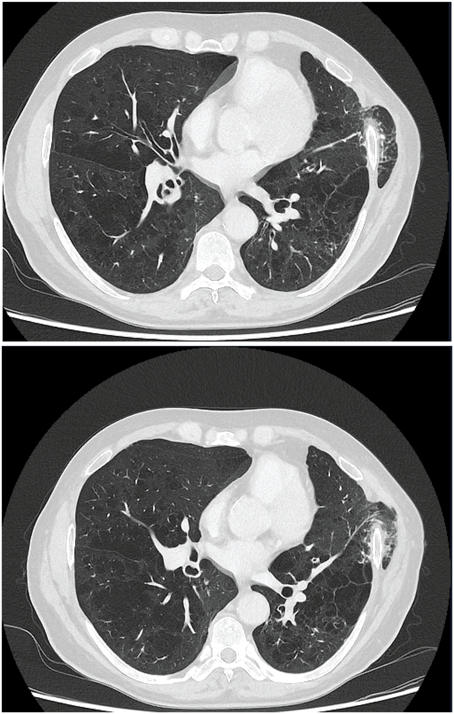
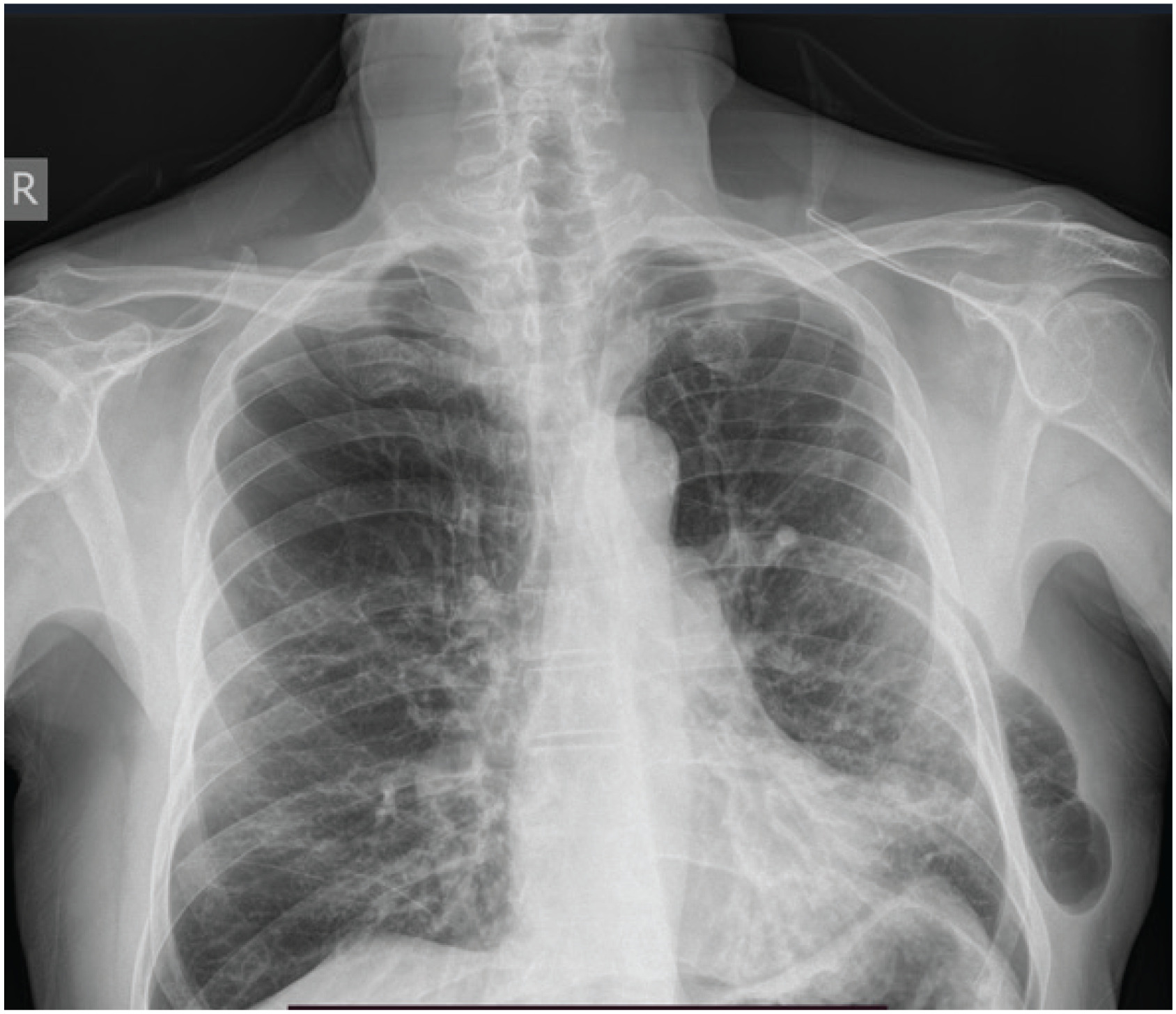
The patient, 67 years old, male, former smoker (50 packets per year), medical history of severe COPD (FEV1=1.22 L, 40% of predicted), came to the emergency department due to fever, dyspnea, and productive cough. The patient’s medical history included segmental resection of the left upper pulmonary lobe for diagnostic purposes, due to the presence of a lung nodule, four years ago. The biopsy indicated a cryptogenic organizing pneumonia. The patient’s diagnosis was infective COPD exacerbation and was treated with antibiotics, methylprednisolone 40 mg iv for 5 days, and bronchodilator therapy. Upon physical examination, a small protrusion was noted in the left anterior chest wall, which became more prominent during coughing and deep breathing. He underwent a chest CT scan, which revealed severe emphysema in all lung zones, as well as a lung hernia to the left upper lung zone (Figure 1), additionally an X-ray was performed (Figure 2). Although, the patient did not report any symptoms, associated with lung hernia, a future surgical intervention was decided.
DISCUSSION
Lung hernia is a rare health condition. Lung hernias occur when the lung parenchyma along with pleural membranes protrude through a defect in a chest wall. Classification is based on both the etiology (congenital and acquired) and anatomic location (cervical, thoracic, diaphragmatic, mediastinal). A lung hernia is described after a trauma or after thoracic surgery. Most patients do not describe any symptoms associated with lung hernia. Also, it should be noted that lung hernia typically remains stable in size or progression is very slow1-3. There is controversy as to the ideal management of these patients. A more conservative method, such as observation, could be selected for asymptomatic patients4,5. However, conditions that increase intrathoracic pressure, such as coughing, can exacerbate the symptoms and reveal the hernia, and patients are at risk of developing complications, like strangulation of the hernia contents. So, surgical intervention is recommended for those occasions, with various techniques6.
CONCLUSION
In our case, lung hernia is characterized as thoracic, acquired, due to the prementioned surgical procedure. The patient has a medical history of severe COPD and cough is a prominent symptom. Also, he suffered more than two exacerbations per year. Consequently, there is a greater risk of deterioration of lung hernia, due to increased intrathoracic pressure. Recommended treatment includes surgical intervention with lung hernia reduction and repair of chest wall defect, so he was referred to a thoracic surgeon for specialized management.