


INTRODUCTION
Bacterial pneumonia remains a significant global health burden despite advances in antibiotic therapy, affecting approximately 4 million people annually, with 25% requiring hospitalization1. Parapneumonic pleural effusions (PPE) complicate around 40% of these cases, with 10% necessitating surgical intervention2. PPE is defined as any pleural effusion associated with bacterial pneumonia, lung abscess, or bronchiectasis, progressing through three stages: exudative, fibrinopurulent (Stage II), and organization. The exudative stage is characterized by sterile free-flowing pleural fluid that usually responds well to antibiotic therapy. The fibrinopurulent (Stage II) stage involves fibrin deposition, septation, loculation, and purulent fluid formation, often requiring pleural drainage in addition to antibiotics. In the organizing stage, fibroblast proliferation and pleural thickening result in a fibrous peel that may trap the lung and often necessitates surgical intervention3.
Management of Stage II PPE remains challenging due to fibrin membranes that compartmentalize pleural fluid, impeding effective drainage4. Intrapleural fibrinolytics, such as streptokinase (SK), aim to dissolve these membranes, enhancing fluid clearance and potentially reducing the need for surgery5. Early studies, like Bergh et al.5, reported radiological improvements with SK in empyema patients, while Diacon et al.6 found it safe and effective, reducing surgical referrals6. However, conflicting results, such as those from the MIST1 trial showing no benefit7, underscore the need for further investigation.
The findings may be relevant to resource-limited settings, although further studies are needed to evaluate the economic implications of intrapleural streptokinase.
METHODS
This hospital-based randomized controlled trial (RCT) was conducted in the Department of Respiratory Medicine at a tertiary care center in India from November 2022 to April 2024, following Institutional Ethics Committee approval. Written informed consent was obtained from all participants. The study was prospectively registered with the Clinical Trials Registry–India (CTRI) (Registration No. CTRI/2023/12/060882).
Study population and sample size
Patients aged >18 years with Stage II parapneumonic pleural effusions (PPE), confirmed by pleural fluid analysis (pH <7.0, glucose <40 mg/dL, positive Gram stain or culture) or imaging evidence of multiloculation without frank pus, were included. Exclusion criteria comprised refusal of consent, malignant effusions, trauma-related effusions, hemothorax, frank empyema, hydropneumothorax, pyopneumothorax, bleeding disorders, or anticoagulant use. Based on the Diacon et al.6 success rates (82.0% SK vs 48.0% placebo), a sample size of 60 (30 per group) was calculated using the Fleiss method (α=0.05, β=0.2).
Randomization and intervention
Participants were randomized into two groups using block randomization (Ralloc method). The allocation sequence was generated by the supervising investigator using Ralloc software and concealed using sequentially numbered opaque sealed envelopes. This was an open-label study; therefore, participants, treating physicians, and radiological outcome assessors were aware of treatment allocation. The test group received ICD plus two doses of intrapleural streptokinase (250000 IU, reconstituted in 100 mL normal saline, 24 hours apart), using the commercially available preparation Streptokinase 1.5 million IU vial, manufactured by Cadila Healthcare Ltd., India (diluted appropriately to the required dose), with the tube clamped for 4 hours post-instillation. The control group received standard care (ICD alone), and no placebo instillation was performed because the study was designed to compare adjunctive streptokinase with standard clinical management. Both groups were initially started on empirical antibiotic therapy, which was subsequently modified according to pleural fluid culture and antibiotic susceptibility results, whenever available.
Data collection and outcome measures
Baseline data included demographic variables (age and sex), chief complaints (fever, cough, chest pain, and dyspnea), and comorbidities recorded from the patients’ medical history and clinical records. The comorbidities assessed included diabetes mellitus, hypertension, chronic kidney disease (CKD), coronary heart disease (CHD), and previous history of tuberculosis. Baseline pleural fluid volume was assessed by ultrasonography (USG; Samsung HS70A/RS80A). Chest radiographs (CXR; Siemens Heliophos-D) and USG were performed on Days 1 and 4 to evaluate outcomes. Primary outcomes included lung expansion on chest radiography, assessed on Day 4 by visual estimation of the percentage of re-expansion relative to the contralateral lung by a consultant radiologist as part of routine clinical practice. Lung expansion was categorized as near complete (≥90% re-expansion with minimal residual opacity) or partial (<90% re-expansion with persistent opacity or pleural thickening). Residual pleural collection was assessed by ultrasonography 24 hours after the second streptokinase dose (Day 4) and categorized as no/minimal (no detectable fluid or <50 mL in a single pocket) or moderate/gross (≥50 mL fluid or multiple loculated pockets). The same consultant radiologist performed all radiological assessments using routine clinical interpretation. Secondary outcomes included total and day-wise ICD drainage (mL), duration of ICD placement (days), hospital stay (days), and complications (anaphylaxis, rash, worsened dyspnea, intrapleural bleeding).
Statistical analysis
Categorical variables were summarized as frequencies and percentages, while continuous variables were expressed as mean ± standard deviation (SD). Comparisons between groups were performed using the chi-squared test for categorical variables and the unpaired t-test for continuous variables. Statistical analyses were performed using EPI Info™ version 7.08. A p-value of <0.05 was considered statistically significant.
RESULTS
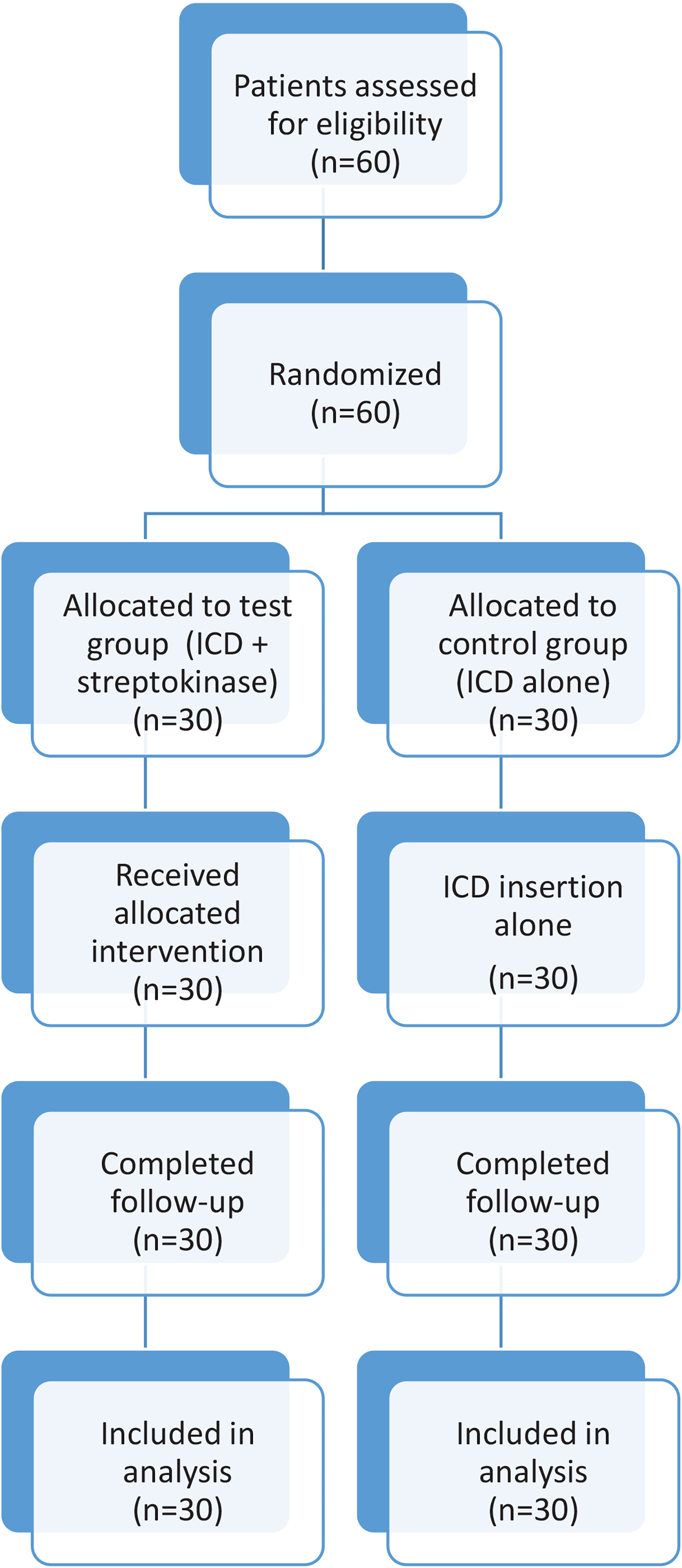
Sixty patients were enrolled, with 30 in each group. Baseline characteristics and outcomes are summarized in Figure 1.
Demographics and comorbidities
Table 1 presents demographics and comorbidities. No significant differences were observed in age or gender (p>0.05), with most patients aged 31–40 years (30.0% in both groups) and a mean age of 42.2 ± 10.12 years. Males predominated in both groups with a male-to-female ratio of 18:12 in the test group and 19:11 in the control group. Diabetes mellitus was the most common comorbidity (16.7% test, 20.0% control), with no significant intergroup differences (p>0.05).
Table 1
Baseline demographic characteristics, comorbidities, and pleural fluid volume of patients with Stage II parapneumonic pleural effusion enrolled in the randomized controlled trial
Fluid volume at diagnosis
Table 1 also shows pleural fluid volume at diagnosis. Gross effusions were more frequent in the test group (56.6%) than the control group (46.6%), but this was not significant (p=0.12).
Radiological outcomes
Table 2 combines lung expansion and residual collection data. Near complete lung expansion was more frequent in the test group (60.0%) than the control group (43.3%), though not significant (p=0.09). Residual collection differed significantly (p=0.004), with minimal/no collection in 86.6% of the test group versus 50.0% of the control group.
Table 2
Comparison of clinical and radiological outcomes between the streptokinase plus intercostal drainage group and the intercostal drainage alone group in patients with Stage II parapneumonic pleural effusion
Drainage and hospitalization
Table 3 presents total drainage, ICD duration, and hospital stay. Total drainage was significantly higher in the test group (e.g. 3648 ± 1250 mL for gross effusions vs 1986 ± 674 mL in controls, p<0.0001). ICD duration was shorter in the test group (7.81 ± 1.85 days) versus controls (11.00 ± 2.26 days, p<0.0001), but hospital stay did not differ significantly (13.92 ± 2.28 vs 14.38 ± 1.33 days, p=0.26).
Table 3
Comparison of treatment-related complications between the streptokinase plus intercostal drainage group and the intercostal drainage alone group
Success rate
Table 4 shows success, defined as no/minimal residual loculations on USG 24 hours post-second SK dose. The test group had a higher success rate (86.6%) than the control group (50.0%, p=0.001). Failures (4 test, 15 control) were referred for video-assisted thoracoscopic surgery (VATS).
DISCUSSION
This randomized controlled trial evaluated the efficacy of intrapleural streptokinase (SK) as an adjunct to intercostal drainage (ICD) in patients with Stage II parapneumonic pleural effusions (PPE). Overall, patients receiving SK with ICD demonstrated improved pleural fluid drainage, reduced residual pleural collections, shorter ICD duration, and a higher treatment success rate compared with ICD alone. However, no significant differences were observed in hospital stay or lung expansion. These findings suggest that intrapleural SK may be a useful adjunct in the management of Stage II PPE.
The improved drainage and reduction in residual pleural collections observed in the SK group are consistent with the fibrinolytic mechanism of streptokinase, which facilitates disruption of fibrin septations and promotes pleural fluid clearance. Similar findings have been reported by Rahman et al.4, Diacon et al.6 and Bouros et al.9, supporting the role of intrapleural fibrinolytic therapy in selected patients with loculated parapneumonic effusions.
The higher success rate in the test group (86.6% vs 50.0%, p=0.001), defined as minimal or no residual loculations on ultrasonography (USG), further supports SK’s efficacy in breaking down pleural septations, consistent with its mechanism of converting plasminogen to plasmin4. This finding echoes the Omar et al.10 observation of improved adhesiolysis with SK. The significant reduction in residual collection (86.6% minimal/no vs 50.0%, p=0.004) likely stems from SK’s ability to target multiloculated effusions, a hallmark of Stage II PPE, enhancing pleural space clearance5. Bergh et al.5 similarly found SK effective in reducing loculations in empyema. However, lung expansion trended better in the test group (60.0% near complete vs 43.3% control) without reaching significance (p=0.09), possibly due to the limited sample size of 60 patients, which may lack power to detect smaller differences in radiological outcomes7. Maskell et al.7 MIST1 trial noted similar non-significant lung expansion benefits, attributing it to variability in effusion complexity.
Hospital stay showed no significant difference (13.92 ± 2.28 vs 14.38 ± 1.33 days, p=0.26), despite shorter ICD duration in the test group. This could result from systemic factors like comorbidities (e.g. diabetes in 16.6% of test group patients) delaying overall recovery, as suggested by Cameron and Davies11. Notably, all four test group failures had diabetes mellitus, potentially indicating impaired healing or immune response affecting outcomes, a hypothesis supported by Falguera et al.12, who linked diabetes to prolonged pleural inflammation. The higher initial gross effusion rate in the test group (56.6% vs 46.6%, p=0.12) might also contribute to prolonged recovery in some cases, though not significant, as larger effusions can complicate resolution despite SK3.
SK’s safety profile was robust, with no major complications like anaphylaxis or bleeding. The transient fever in one patient and brief chest discomfort in two, likely stem from mild inflammatory responses to SK, as reported by Diacon et al.6, resolving without intervention. This contrasts with MIST1’s null findings7, where inconsistent SK dosing and broader patient inclusion (including Stage III effusions) may have diluted efficacy. Our focus on loculated Stage II effusions and standardized dosing (250000 IU) likely explains the positive results. The MIST2 trial demonstrated the potential benefits of combination intrapleural therapy with tissue plasminogen activator (tPA) and DNase13. In the present study, the higher treatment success rate and shorter duration of intercostal drainage in the streptokinase group suggest potential benefits in resource utilization. However, as treatment costs and overall healthcare expenditure were not directly assessed, the cost-effectiveness of streptokinase cannot be established from the present study.
Limitations
This study has several limitations. Its single-center design and relatively small sample size may limit the generalizability of the findings. In addition, the open-label design may have introduced observer bias, particularly for imaging-based outcomes, as radiological assessments were not blinded. Although baseline characteristics were comparable between the groups, the possibility of residual confounding cannot be excluded. Furthermore, severe cases were excluded, which may limit the applicability of the findings to all patients with parapneumonic pleural effusions. Future research should optimize SK timing, explore adjunctive therapies, and further evaluate its role in Stage II PPE.
CONCLUSIONS
Intrapleural streptokinase as an adjunct to intercostal drainage may improve pleural fluid drainage, reduce residual pleural collections, shorten ICD duration, and increase treatment success in patients with Stage II parapneumonic pleural effusions. However, no significant benefits were observed with respect to hospital stay or lung expansion. These findings suggest that intrapleural streptokinase may be a useful adjunct in selected patients, although the results should be interpreted in light of the study’s single-center design, relatively small sample size, and open-label methodology. Larger multicenter studies are warranted to confirm these findings and further define the role of intrapleural streptokinase in the management of Stage II parapneumonic pleural effusions.