


INTRODUCTION
Chronic obstructive pulmonary disease (COPD) is a chronic, nonspecific inflammatory airway disease characterized by persistent and progressively worsening airflow obstruction and destruction of alveolar structure1. Although it is currently recognized that the primary pathogenic microorganisms responsible for acute exacerbations of chronic obstructive pulmonary disease (AECOPD) are bacteria and viruses, a growing body of research indicates that AECOPD is frequently complicated by invasive pulmonary aspergillosis (IPA)2,3, which is associated with a high case-fatality rate. Although lung biopsy (e.g. transbronchial or surgical lung biopsy) remains the gold standard for diagnosing IPA4, its feasibility is limited by impaired lung function in patients with COPD and low patient acceptance of the procedure. Therefore, this study favors the use of bronchoscopic alveolar lavage sampling as a safer and more feasible alternative. Microbial culture is a core method for pathogen diagnosis5. It offers the advantages of ease of use and low cost, making it one of the most commonly used diagnostic methods in clinical practice at present5,6; however, it is time-consuming, has low sensitivity, and can easily delay the treatment window.
Microbiological rapid on-site evaluation (M-ROSE) is primarily used in clinical practice for the rapid bedside diagnosis of the etiology of lower respiratory tract infections7 and provides immediate morphological evidence, significantly shortening the time to diagnosis. A study has shown that serum inflammatory markers – C-reactive protein (CRP), erythrocyte sedimentation rate (ESR), lactate dehydrogenase (LDH), and procalcitonin (PCT) – have diagnostic value for COPD complicated by IPA8. Combining M-ROSE with serum inflammatory markers and fungal serological markers [galactomannan test (GM test) and (1,3)-β-D-glucan test (G test)]9 not only expands its clinical utility but also holds promise for further improving the diagnostic accuracy of IPA in patients with COPD.
CRP and PCT are widely used as markers of bacterial infection and systemic inflammation. ESR is a traditional marker of systemic inflammation. LDH levels rise during severe pulmonary infections and may be associated with tissue damage. Therefore, this study selected these markers as candidate serum inflammatory markers. These markers can be routinely measured in most clinical laboratories and can be efficiently and cost-effectively incorporated into the diagnostic process for COPD complicated by IPA. This study employed a prospective paired design, using the same bronchoalveolar lavage fluid (BALF) sample to perform both M-ROSE testing and conventional fungal culture simultaneously. With conventional fungal culture as the reference standard, the study evaluated the diagnostic performance and time efficiency of M-ROSE for COPD complicated by IPA, and explored whether combining it with serum inflammatory markers could further improve diagnostic accuracy.
METHODS
Study design, setting, and participants
This study employed a paired-test design and was a prospective cross-sectional diagnostic accuracy study conducted at Huaibei City People’s Hospital from June 2023 to April 2026. It enrolled 108 patients diagnosed with chronic obstructive pulmonary disease (COPD) and suspected of having invasive pulmonary aspergillosis (IPA); all patients were admitted to the Department of Respiratory and Critical Care Medicine.
Inclusion criteria
Based on the consensus criteria of the European Organization for Research and Treatment of Cancer/Mycoses Study Group Education and Research Consortium (EORTC/MSGERC)10, the inclusion criteria were : 1) clinical manifestations suggestive of IPA (e.g. fever, persistent cough, chest pain, hemoptysis, etc.); 2) imaging findings consistent with IPA, chest CT shows pulmonary infiltrates, nodules, or cavities; and 3) ability to tolerate bronchoscopy: Patients who are hemodynamically stable, have an SpO2 >90%, and have no uncontrolled arrhythmias or severe coagulation disorders.
Exclusion criteria
The exclusion criteria were: 1) active massive hemoptysis (hemoptysis volume >100 mL within 24 hours); 2) inability to tolerate bronchoscopy; 3) concurrent neutropenia, malignancy, dialysis, or hematopoietic stem cell transplantation; and 4) concurrent fungal infections or prior use of antifungal medications.
Ethics
This study was reviewed and approved by the hospital’s Ethics Committee (Approval No. 2023–014). All patients or their legal guardians signed written informed consent forms, and no additional medical expenses were incurred as a result of this study.
Specimen collection and laboratory procedures
Specimen collection
Bronchoalveolar lavage fluid (BALF) was collected in accordance with the standard operating procedures recommended in the Chinese Expert Consensus on Cytomorphological Examination of Conventional Bronchial Pathogens (2023)11.
M-ROSE testing
Specimens were centrifuged at 300g for 10 min; the pellet was resuspended in 0.5 mL of saline and stained with the Wright-Giemsa stain. After natural fixation, the specimens were examined under a fluorescence microscope; samples exhibiting specific fluorescent signals consistent with aspergillus characteristics were classified as fungal-positive.
BALF fungal smear and culture
The same BALF specimen was sent to the microbiology laboratory for conventional fungal culture, strictly following the National Clinical Laboratory Operating Procedures12.
Serological testing
Fasting venous blood samples were collected simultaneously and tested in the hospital’s clinical laboratory. CRP was measured using an immunoturbidimetric assay, ESR by the Westergren method, LDH by an enzyme-rate assay, and PCT by a chemiluminescence assay. All tests were performed in accordance with standardized operating procedures on a Roche Cobas 8000 analyzer (Roche Diagnostics, Mannheim, Germany) for CRP, LDH, and PCT, and on a Mindray BC-5180 (Mindray, Shenzhen, China) for ESR.
Interpretation and evaluation of results
M-ROSE on-site image review criteria
M-ROSE test results are independently reviewed on-site using a double-blind method by one pulmonologist and one clinical laboratory physician from this hospital, who then record their interpretations.
M-ROSE positive determination criteria
If a clear, yellow-green, specific fluorescent signal appears in the field of view and its morphology matches the characteristics of aspergillus hyphae or spores, and both physicians confirm this consistently, the result is deemed M-ROSE positive; otherwise, it is negative. When the M-ROSE smear result matches the official laboratory report issued subsequently by the Department of Laboratory Medicine, it is recorded as a diagnostic concordance. The results from the clinical laboratory are used as the reference standard. The diagnostic performance of the M-ROSE smear is calculated using the following formulas:
Sensitivity = number of true positives/(number of true positives + number of false negatives) × 100%;
Specificity = number of true negatives/(number of true negatives + number of false positives) × 100%; and
Diagnostic agreement rate = (number of true positives + number of true negatives)/total number of cases × 100%.
Statistical analysis
SPSS 27.0 (IBM Corp., Armonk, NY, USA)13 and GraphPad Prism 10.1.2 software14 (GraphPad Software, Boston, MA, USA) were used for statistical analysis and graphing. Conventional fungal culture was used as the reference standard to calculate the diagnostic performance metrics of M-ROSE, with 95% confidence intervals (CIs) calculated using the Wilson method. The kappa test was used to assess agreement. Since these measurement data were all collected from the same group of patients, the Friedman test was used to compare the turnaround times for the M-ROSE test, fungal smear, and fungal culture. Serum inflammatory markers and combined diagnostic performance were evaluated using receiver operating characteristic (ROC) curves, and the optimal cutoff value was determined based on the Youden index. A p<0.05 was considered statistically significant.
RESULTS
Baseline characteristics of patients
This study included a total of 108 patients with COPD and suspected IPA. Based on fungal culture results, they were divided into two groups: 92 patients (85.2%) in the fungal culture-positive group and 16 patients (14.8%) in the fungal culture-negative group. There were no statistically significant differences between the two groups in terms of age, BMI, smoking history, or major comorbidities (all p>0.05), suggesting that the baseline data of the two groups were comparable (Table 1).
Table 1
Baseline characteristics of patients with COPD and suspected invasive pulmonary aspergillosis at Huaibei People’s Hospital, June 2023–April 2026 (N=108)
[i] Patients were divided into positive and negative groups based on fungal culture results. For the p-values, Fisher’s exact test was used for cerebrovascular disease, diabetes, liver disease, endocrine system diseases, digestive system diseases, malignant tumors, tuberculosis, and malnutrition. The Pearson chi-squared test was used for all other conditions.
Evaluation of detection results and diagnostic performance of M-ROSE compared to conventional fungal culture as the reference standard
The overall diagnostic performance metrics of M-ROSE are detailed in Table 2. The kappa consistency test showed that the overall agreement rate between the two methods was 87.04%, with a kappa value of 0.768 (p<0.001), indicating a high degree of agreement between M-ROSE and conventional fungal culture. A comparison of the specific test results for the two methods is presented in Table 2.
Table 2
A comparison of M-ROSE and conventional fungal culture results in patients with chronic obstructive pulmonary disease (COPD) and suspected invasive aspergillosis admitted to Huaibei Municipal People’s Hospital, June 2023–April 2026 (N=108)
| Positive fungal culture (N=92) n (%) | Fungal culture was negative (N=16) n (%) | Total | |
|---|---|---|---|
| M-ROSE positive | 82 (89.13) | 4 (25.00) | 86 |
| M-ROSE negative | 10 (10.87) | 12 (75.00) | 22 |
| Total | 92 (100) | 16 (100) | 108 |
Comparison of time efficiency between M-ROSE and conventional fungal culture
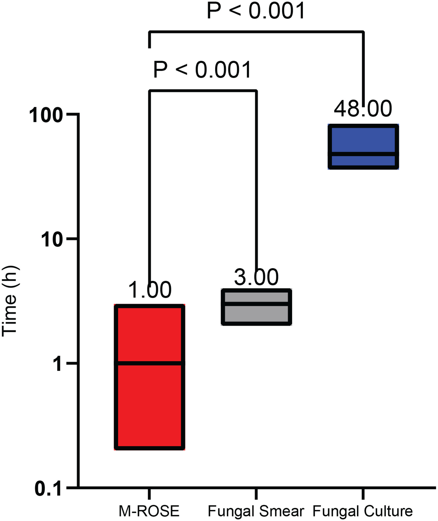
Figure 1 shows the results of a comparison of turnaround times for M-ROSE, fungal smears, and fungal cultures. M-ROSE had the shortest median turnaround time, while fungal culture had the longest. The Friedman test revealed that the differences in turnaround times among the three methods were statistically significant (p<0.001).
Analysis of the diagnostic performance of individual and combined serum inflammatory markers with M-ROSE
Diagnostic performance of individual serum markers
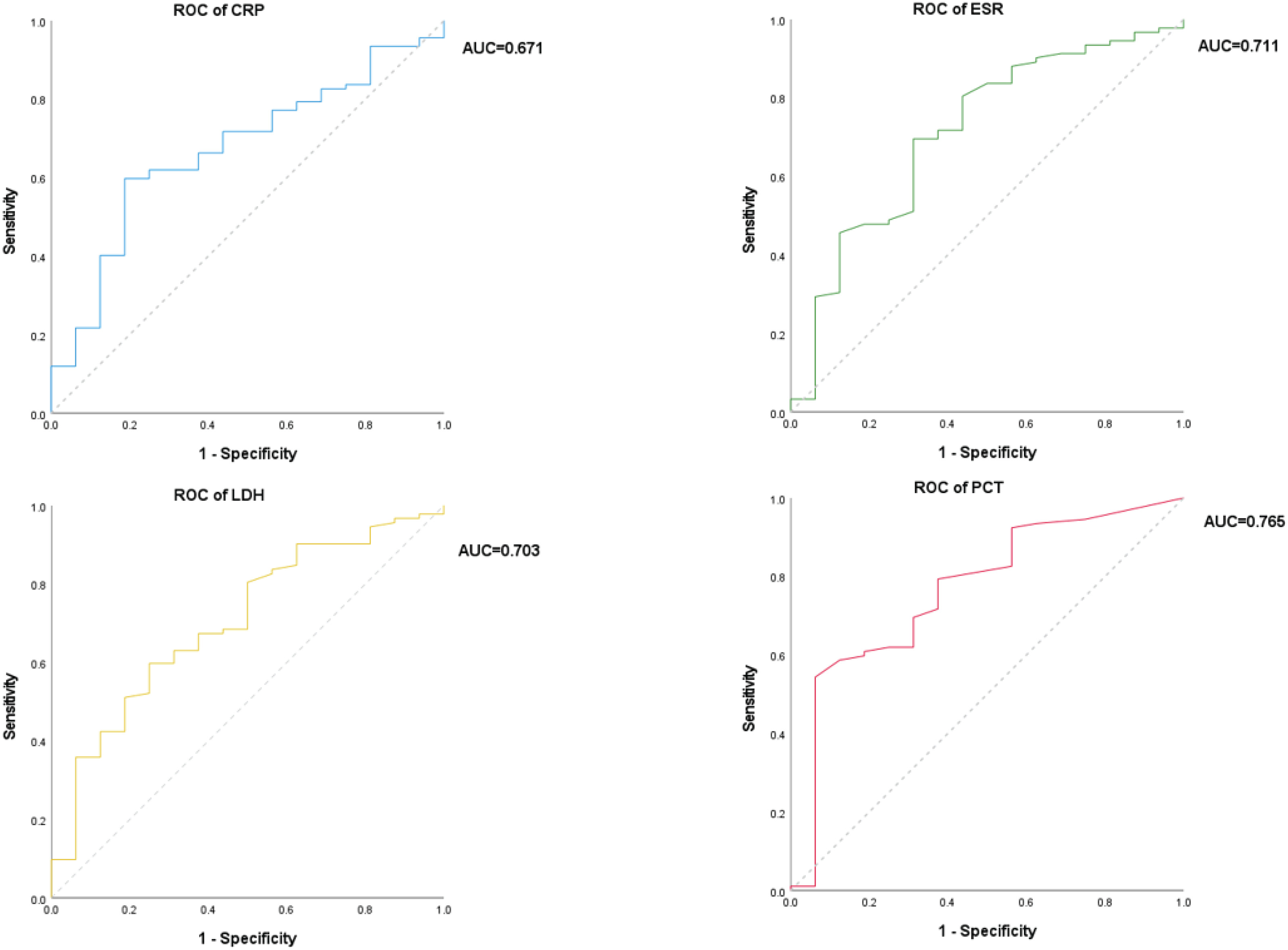
Based on the optimal diagnostic cutoff values calculated from the data in this study (CRP >30.8 mg/L, ESR >35.9 mm/h, LDH >238 U/L, PCT >0.21 ng/mL), the diagnostic performance of each serum inflammatory marker in individually diagnosing COPD complicated by IPA was evaluated. ROC curve analysis showed (Figure 2) that the area under the curve (AUC) for the four serum inflammatory markers were as follows: CRP 0.671 (p=0.030), ESR 0.711 (p=0.007), LDH 0.703 (p=0.010), PCT 0.765 (p=0.001), with PCT demonstrating the highest relative diagnostic performance. The diagnostic performance of each individual marker was lower than that of the combined diagnostic model.
Figure 2
ROC curves for the use of serum inflammatory markers (CRP, ESR, LDH, and PCT) in the diagnosis of patients with suspected invasive aspergillosis in the setting of chronic obstructive pulmonary disease (COPD) at Huaibei Municipal People’s Hospital from June 2023 to April 2026 (N=108)
Diagnostic performance of M-ROSE combined with four inflammatory markers
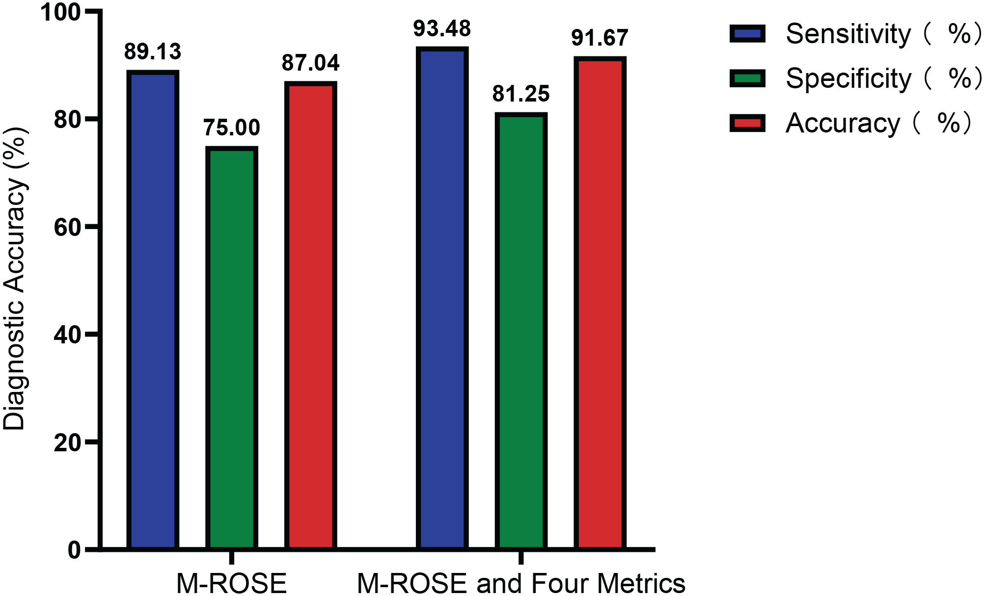
Compared with M-ROSE used alone (Table 3), the diagnostic performance of M-ROSE combined with four inflammatory markers for COPD with IPA showed improvements in sensitivity, specificity, and overall agreement rate, suggesting that the combined approach has higher diagnostic performance (Table 4).
Table 3
Diagnostic performance of M-ROSE testing for invasive aspergillosis in patients with suspected IPA among those with chronic obstructive pulmonary disease (COPD) admitted to Huaibei Municipal People’s Hospital, June 2023–April 2026 (N=108)
| Indicators | % | 95% CI |
|---|---|---|
| Sensitivity | 89.13 | 80.92–94.66 |
| Specificity | 75.00 | 47.62–92.73 |
| Positive predictive value | 95.35 | 89.74–97.96 |
| Negative predictive value | 54.55 | 38.52–69.68 |
| Accuracy | 87.04 | 79.21–92.73 |
| Kappa | 0.768 | |
| p | <0.001 |
Table 4
Diagnostic performance of M-ROSE combined with serum inflammatory markers (CRP, ESR, LDH, PCT) in patients with suspected invasive aspergillosis overlying chronic obstructive pulmonary disease (COPD) admitted to Huaibei Municipal People’s Hospital, June 2023–April 2026 (N=108)
| Indicators | % | 95 % CI |
|---|---|---|
| Sensitivity | 93.48 | 86.34–97.57 |
| Specificity | 81.25 | 54.35–95.95 |
| Positive predictive value | 96.63 | 91.17–98.76 |
| Negative predictive value | 68.42 | 49.11–82.95 |
| Accuracy | 91.67 | 84.77–96.12 |
| Kappa | 0.694 | |
| p | <0.001 |
[i] A comparison of the performance of M-ROSE used alone versus the combined approach is shown in Figure 3.
Figure 3
A comparison of the diagnostic performance of M-ROSE testing for invasive aspergillosis in patients with chronic obstructive pulmonary disease (COPD) at Huaibei Municipal People’s Hospital and the combination of M-ROSE testing with serum inflammatory markers (CRP, ESR, LDH, PCT), June 2023–April 2026 (N=108)
DISCUSSION
The results of this study indicate that: 1) M-ROSE has a sensitivity of 89.13% and a specificity of 75.00% for the rapid detection of patients with suspected IPA associated with COPD; compared with conventional fungal culture, the overall agreement rate was 87.04% (κ=0.768). 2) The testing time for M-ROSE is shorter than that for fungal culture and fungal smear. 3) Serum inflammatory markers (CRP, ESR, LDH, PCT) have low diagnostic performance when used alone; and 4) When M-ROSE is combined with serum inflammatory markers, diagnostic performance is further improved, with a sensitivity of 93.48%, a specificity of 81.25%, and an accuracy of 91.67%.
Clinical methods for diagnosing pulmonary aspergillosis are diverse: chest X-rays lack specificity, showing patchy pulmonary infiltrates and ground-glass opacities15; some patients may present with nodular or consolidation shadows. Since these findings are similar to those of bacterial pneumonia, it is difficult to distinguish between the two using routine imaging studies. Consequently, some patients are misdiagnosed with bacterial pneumonia; after receiving symptomatic treatment such as antimicrobial therapy, their condition shows no improvement, delaying the optimal timing for antifungal treatment. In immunocompromised patients, a study by Sarrafzadeh et al.16 reported that serum GM testing had a sensitivity of 69.2% and a specificity of 72.2% for the diagnosis of IPA, indicating moderate diagnostic performance overall; a study by Niu et al.17 noted that the positive rate for aspergillus culture was only 29.0% in patients with acute exacerbations of COPD complicated by IPA. The EORTC/MSGERC (2020) guidelines recommended the use of PCR technology for IPA diagnosis for the first time10. Gene sequencing technologies (such as tNGS and mNGS), which have evolved from PCR, have made significant progress and can simultaneously detect ten to hundreds of known pathogenic microorganisms. However, these technologies currently have shortcomings, such as high testing costs and the need for specialized teams to interpret results, and therefore cannot yet serve as routine diagnostic tools in primary care settings. Rapid On-Site Evaluation (ROSE) primarily includes cytological rapid on-site evaluation (C-ROSE) and M-ROSE18. The core clinical value of M-ROSE is primarily reflected in three aspects: 1) For fungi such as candida and aspergillus, preliminary identification can be made based on morphological characteristics; 2) Repeated specimen collection at the site ensures that specimen quality is optimized as much as possible; and 3) Under a microscope, observations of cell classification, counting, and proportions allow for a rough distinction between infectious and non-infectious diseases, thereby enabling the formulation of preliminary treatment strategies19. Compared with other fungal assessment methods, M-ROSE can rapidly obtain morphological information from lower respiratory tract specimens at the bedside, providing a basis for preliminary judgment of fungal infections18.
Using conventional fungal culture as the reference standard, this study evaluated the diagnostic performance of M-ROSE in patients with COPD complicated by IPA. The results showed that M-ROSE had a sensitivity of 89.13%, a specificity of 75.00%, and an overall agreement rate of 87.04%, indicating a high degree of consistency between M-ROSE and the reference standard. These findings are consistent with those reported by Shi et al.20. It is worth noting that the negative predictive value of M-ROSE was only 54.55%, suggesting that a relatively high proportion of fungal cultures remains positive even when M-ROSE yields a negative result. This may be related to factors such as low fungal load in the BALF of some patients, suboptimal sampling sites, or insufficient operator experience. Traditional microbial culture requires 3–5 days and has a relatively low positivity rate, whereas the M-ROSE technique can significantly shorten the testing time. Tao et al. 7 reported that M-ROSE results were reported 3.61 ± 1.28 days faster than pathogen culture and 1.64 ± 1.07 days faster than mNGS; Hong et al.21 found that the median time to report ROSE results was only approximately 8.0 minutes, whereas traditional pathology reports took 5645.5 minutes. This study is consistent with the above findings: the median reporting time for M-ROSE (1.00 hour) was significantly shorter than that for fungal culture (48.00 hours), further validating the rapid diagnostic advantages of ROSE technology. In recent years, metagenomic next-generation sequencing (mNGS) has demonstrated high sensitivity in the diagnosis of IPA. A study by Niu et al.17 on patients with AECOPD complicated by IPA showed that BALF mNGS had a sensitivity of 70.9%, which was significantly higher than that of fungal culture (29.0%) and the GM test (41.9%). A multicenter study by Zhong et al.22 also confirmed that the sensitivity of BALF mNGS can reach 81.9%. The aforementioned studies also pointed out limitations of mNGS, including high testing costs, inconsistent interpretation criteria, and a turnaround time of 24–48 hours; they explicitly stated that mNGS should serve as a complementary tool to traditional testing rather than a replacement. Therefore, in primary-care hospitals where mNGS is not yet widely available, the M-ROSE combined protocol can serve as a practical method for early rapid screening and initiating treatment.
Further analysis showed that serum inflammatory markers (CRP, ESR, LDH, PCT) alone have limited diagnostic utility for COPD complicated by IPA; ROC curve analysis indicated that PCT had the highest relative diagnostic utility, with an AUC of 0.765. When M-ROSE was used in combination with the aforementioned four serum inflammatory markers, diagnostic performance improved significantly: sensitivity increased to 93.48%, specificity to 81.25%, and accuracy to 91.67%, all of which were superior to the results obtained with M-ROSE alone. These results suggest that serum inflammatory markers alone lack specificity and are prone to interference from other bacterial infections; whereas M-ROSE can directly identify aspergillus at the pathogen level.
Limitations
This study has certain limitations, primarily reflected in the following: 1) We acknowledge that conventional fungal culture is not a true gold standard for IPA, as a positive culture may reflect airway colonization rather than invasive disease, and a negative culture does not exclude IPA due to limited sensitivity. Therefore, we refer to it as the reference standard in this study; 2) As a single-center, small-sample-size prospective analysis, the study’s conclusions are subject to certain limitations. Future large-scale, multicenter prospective studies are needed to further validate the diagnostic efficacy of the combination of M-ROSE and serum inflammatory markers in patients with COPD complicated by IPA; 3) The interpretation of M-ROSE results depends on the operator’s experience, and subjective differences may exist among different interpreters; currently, there is a lack of unified, standardized operating procedures and interpretation criteria; and 4) The optimal diagnostic thresholds for serum inflammatory markers (CRP, ESR, LDH, PCT) in this study were calculated based on the data from this cohort; the applicability of these thresholds in different populations and on different testing platforms requires external validation.
CONCLUSIONS
Tthe combination of M-ROSE technology and serum inflammatory markers holds promise for the rapid diagnosis of fungal infections and may aid in early clinical decision-making. However, due to the limitations of the reference standard and the single-center study design, the findings of this study are preliminary. Further validation through large, multicenter studies using composite reference standards is required before recommending its use in routine clinical practice.