


INTRODUCTION
Pulmonary siderosis, also known as welder’s lung, is an occupational lung disease caused by chronic inhalation of iron oxide dust or fumes1,2. Traditionally considered a benign pneumoconiosis without fibrosis, recent evidence suggests that prolonged or mixed-dust exposure may result in interstitial lung disease3-5. Radiologically, pulmonary siderosis may present as diffuse micronodules, occasionally mimicking miliary tuberculosis, particularly in TB-endemic countries6,7. Bronchoalveolar lavage (BAL) with Prussian blue staining offers a simple, cost-effective, and confirmatory diagnostic approach8. This case is unique due to its striking miliary radiological pattern in a patient with prior extrapulmonary tuberculosis, creating a significant diagnostic dilemma.
CASE REPORT
A 51-year-old man presented to the respiratory outpatient department with cough producing mucoid expectoration for 10 days, associated with low-grade evening fever for 5–6 days and mild exertional dyspnea (Modified Medical Research Council Grade I, static for one month). He denied hemoptysis, chest pain, palpitations or orthopnea. He had worked in a stainless-steel factory for several years but had ceased exposure two years prior and reported no ongoing or alternative iron exposure. He was currently employed as a security guard. He had been treated for extrapulmonary tuberculosis (cervical lymphadenopathy) eight months earlier, with no documented pulmonary involvement at that time for which he completed a full six-month course of antitubercular therapy. There was no history of diabetes, hypertension, ischemic heart disease, or occupational exposure to silica, asbestos, or organic dusts. He denied any history of TB contact or smoking. On examination, he was afebrile and hemodynamically stable. Oxygen saturation was 97% on room air. Bilateral diffuse fine inspiratory crackles were heard over the chest. There was no cyanosis, clubbing, or pedal edema. Routine hematological and biochemical investigations were within normal limits.
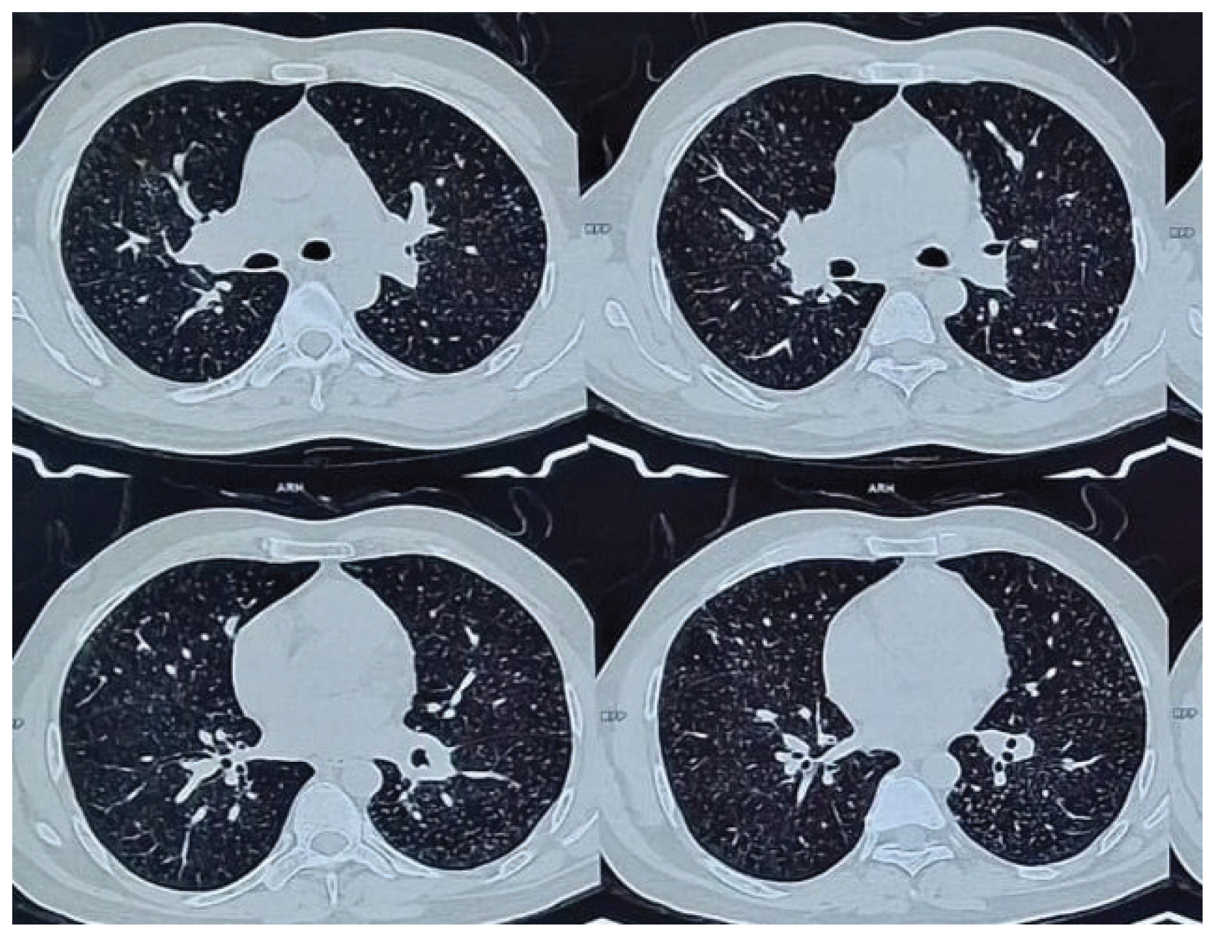
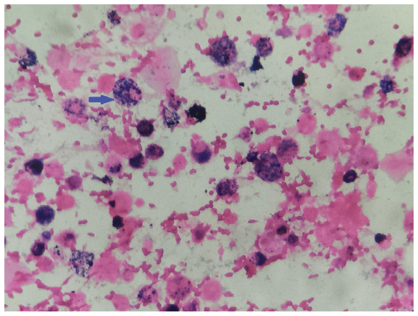
Chest radiograph showed multiple tiny diffuse nodular opacities in both lung fields. HRCT thorax showed diffuse miliary nodules involving all lobes without fibrosis or cavitation (Figure 1). BAL analysis was negative for AFB smear, Lowenstein–Jensen culture, MGIT culture, bacterial and fungal cultures, and malignant cells. Prussian blue staining demonstrated numerous hemosiderin-laden macrophages (Figure 2). The patient was counseled to avoid any further exposure to iron dust and was advised to use protective respiratory equipment at work. The patient received inhaled bronchodilators, formoterol (4.8 μg) with glycopyronium (9 μg) twice a day for 1 month and supportive care. At follow-up at 3 months, symptoms improved and imaging showed no progression.
DISCUSSION
Pulmonary siderosis results from occupational inhalation of iron oxide particles1-4. Although historically considered benign, persistent radiological abnormalities may remain long after exposure cessation5. However, modern HRCT and histopathological studies indicate that some patients may develop interstitial fibrosis, particularly with concurrent exposure to silica or other irritants2,5. Inhaled iron particles are engulfed by alveolar macrophages and deposited as hemosiderin within alveoli and interstitium. These iron-laden macrophages can be demonstrated by the Prussian blue reaction, which stains ferric iron as deep blue granules3. Persistent exposure triggers macrophage activation and release of pro-fibrotic cytokines, contributing to interstitial changes5.
Most cases are asymptomatic and detected incidentally on imaging. Symptomatic patients may present with chronic cough or exertional dyspnea. In our patient, respiratory symptoms appeared after cessation of exposure, suggesting a delayed inflammatory response possibly potentiated by recent infection. Chest radiographs typically show fine nodular opacities predominantly in the upper lobes. HRCT provides greater sensitivity, demonstrating micronodules, ground-glass opacities, or reticulation6. Pulmonary siderosis may mimic miliary tuberculosis, emphasizing the need for tissue or cytological confirmation7. Cases with diffuse deposition of iron particles and miliary patterns have been rarely described, especially in workers with prolonged exposure and fine-particle inhalation1,8. Importantly, in our patient, co-existent miliary tuberculosis was carefully excluded through repeated microbiological evaluation and radiological stability on follow-up. Although coexistence has been reported, negative cultures and absence of progression argued against active TB6,7.
Bronchoalveolar lavage is a minimally invasive diagnostic method when transbronchial or surgical biopsy is not indicated. Demonstration of hemosiderin-laden macrophages positive for Prussian blue stain confirms the presence of intra-alveolar iron3. Differentials include idiopathic pulmonary hemosiderosis, chronic alveolar hemorrhage, and mixed-dust pneumoconiosis; clinical and occupational history aids differentiation4. There is no specific pharmacological therapy for pulmonary siderosis. The mainstay is elimination of exposure to iron oxide dust. Symptomatic management includes bronchodilators and, occasionally, corticosteroids if airway inflammation predominates. Prognosis can be good in pure siderosis and sometimes regression of radiological findings after cessation of exposure have been documented5,9.
CONCLUSION
Pulmonary siderosis can closely mimic miliary tuberculosis, particularly in TB-endemic settings. This case highlights the critical importance of occupational history and targeted diagnostic evaluation. BAL with Prussian blue staining can prevent misdiagnosis and unnecessary antitubercular therapy.