



INTRODUCTION
Pulmonary imaging research is advancing rapidly, driven by unmet diagnostic needs and technological innovation1-3. Advances in technology and artificial intelligence (AI) promise to transform respiratory disease diagnosis and management4-6. Among these, thoracic ultrasound (TUS) has profoundly reshaped not only pleural disease management but also the assessment of parenchymal pathology, pneumothorax detection, and peri-procedural guidance over recent decades7,8. However, conventional ultrasound remains highly operator-dependent, with subjective interpretation, substantial inter-observer variability, and predominantly anatomical information, providing limited functional or molecular insight. These constraints have fueled growing interest in advanced imaging strategies and AI-based applications designed to broaden the diagnostic scope of TUS beyond conventional assessment9. In this narrative review, we critically introduce and appraise several of the most promising investigational thoracic ultrasound approaches, summarizing their current evidence and the key challenges that will determine whether – and to what extent – they may inform and integrate into routine clinical practice. These domains were selected based on the availability of published clinical evidence, their direct relevance to interventional pulmonology and pleural disease practice, and their potential to extend diagnostic capabilities beyond conventional thoracic ultrasound assessment. Techniques discussed include advanced M-mode–based functional assessment for non-expandable lung prediction, color Doppler ultrasound for pre-procedural intercostal vascular mapping, quantitative pixel-based image analysis for pleural effusion characterization, ultrasound elastography for pleural stiffness evaluation and biopsy targeting, contrast-enhanced ultrasound for optimization of pleural lesion sampling, and AI-driven applications for image acquisition, interpretation, and diagnostic support. A summary of these emerging investigational techniques, including their primary applications, key strengths, main limitations, and current clinical status, is provided in Table 1 and detailed in the following sections.
Table 1
Summary of emerging investigational thoracic ultrasound techniques and artificial intelligence applications
This narrative review was conducted through a structured literature search of PubMed, Embase, and Scopus, from inception to February 2026. Search terms encompassed combinations of the following: ‘thoracic ultrasound’, ‘pleural ultrasound’, ‘M-mode’, ‘color Doppler’, ‘quantitative pixel analysis’, ‘elastography’, ‘contrast-enhanced ultrasound’, ‘CEUS’, and ‘artificial intelligence’, applied individually and in combination for each technique reviewed. Studies were eligible for inclusion if they reported original clinical data, systematic reviews, meta-analyses, or relevant guidelines pertaining to the investigational applications of TUS addressed in this review. Given the narrative nature of this work, no formal PRISMA-compliant screening process was applied; study selection was guided by clinical relevance, methodological quality, and recency of publication, with priority given to prospective designs, multicenter studies, and peer-reviewed publications in respiratory medicine and ultrasound journals.
COMMENTARY
M-mode ultrasound: investigating non-expandable lung prediction
M-mode (motion mode) ultrasound provides a one-dimensional representation of tissue movement along a single ultrasound beam over time. In patients with pleural effusion and lung collapse, it may show subtle pleural or lung motion during breath-hold, including cardiopulmonary-related movements. It has been explored as a possible adjunct for assessing lung mobility and estimating the risk of non-expandable lung before pleural procedures, but evidence remains limited and not yet validated.
Non-expandable lung affects approximately 30% of malignant pleural effusions, significantly impacting management decisions10. Early pre-procedural identification could guide optimal intervention selection, avoiding ineffective pleurodesis attempts and facilitating timely indwelling catheter placement. M-mode ultrasound during breath-hold has been investigated for this purpose by measuring displacement of collapsed lung over cardiac cycles.
Initial studies reported promising results. A prospective study of 81 patients found M-mode achieved an area under the curve (AUC) of 0.79 for predicting non-expandability, with lung displacement <0.8–1.2 mm suggesting non-expandability and guiding intervention choice11.
A subsequent prospective study of 49 patients confirmed M-mode as the best-performing simple technique (AUC=0.81), superior to B-mode and substantially better than shear wave elastography12.
Methodological concerns include small sample sizes, predominance of single-center designs, absence of standardized measurement protocols regarding probe positioning and breath-hold technique, and limited inter-observer reliability data.
While M-mode assessment represents an accessible, radiation-free approach requiring only standard ultrasound equipment and worthy of continued investigation, current evidence is insufficient to support routine clinical implementation13.
From a clinical perspective, the moderate discriminative capacity reflected by AUC values of 0.79–0.81 must be contextualized by several important factors. No prospective outcome data have yet demonstrated that M-mode-guided management decisions translate into improved clinical outcomes. The displacement threshold reported to indicate non-expandability varies between studies (0.8–1.2 mm), reflecting underlying methodological heterogeneity rather than a biologically validated cutoff. Inter-observer reliability has not been formally evaluated in multicenter settings, limiting confidence in the reproducibility of measurements across operators and institutions.
Color Doppler intercostal artery screening: pre-procedural vascular mapping
Color Doppler ultrasound detects blood flow by measuring frequency shifts in reflected ultrasound waves, color-coding the direction and relative velocity of flow in real time. Applied to the thoracic wall prior to pleural procedures, it may enable identification of intercostal arteries within the intended puncture zone, providing anatomical information that complements conventional B-mode guidance.
Hemorrhagic complications from intercostal artery injury, though uncommon, represent serious procedural risks. The intercostal artery exhibits considerable anatomical variability, particularly in elderly patients and upper intercostal spaces, with aberrant courses documented in cadaveric studies14. Color Doppler ultrasound enables pre-procedural vascular mapping to identify vessels and guide safer puncture site selection.
A prospective study of 596 pleural procedures attempted screening in 95% of cases, successfully identifying vessels in 53%15. When visualized, operators modified the puncture site in 30% of these cases (16% of all procedures). The complication rate was 0.17%.
The DIVOT (Doppler Identification of Vessels On Thoracentesis) protocol has been proposed, providing a structured scanning methodology requiring approximately 2 minutes additional time16.
However, critical evidence gaps remain. No study has demonstrated that Doppler screening reduces bleeding complications compared to standard anatomical approaches – the ultimate measure of clinical utility. The observed low complication rate may reflect careful technique rather than screening benefit. Detection varies substantially with probe type, operator experience, and body habitus. Crucially, non-visualization does not guarantee vessel absence, potentially creating false reassurance. The 2023 British Thoracic Society Clinical Statement explicitly identifies this as a research priority, questioning whether Doppler reduces bleeding risk17. While technically feasible and theoretically attractive as a safety adjunct, current evidence is insufficient to recommend routine implementation.
Quantitative image analysis: pixel-based effusion characterization
Conventional ultrasound assessment of pleural effusions relies on subjective visual interpretation. Qualitative features such as septations, fibrin strands, and increased echogenicity are more frequently observed in exudative effusions; however, their overall diagnostic performance is inconsistent and insufficient to support clinical decision-making when used in isolation. To overcome these limitations, quantitative pixel density analysis has been explored to provide more objective assessment18 (Figure 1).
Figure 1
Illustrative example of quantitative pixel-based echogenicity analysis applied to a pleural effusion using ImageJ software (National Institutes of Health, USA). The manually delineated region of interest (ROI), clearly labelled within the effusion on the ultrasound image, is used to measure mean pixel intensity as a quantitative surrogate of fluid echogenicity. This figure is provided as a didactic illustration of the analytical workflow only; it does not represent a formal diagnostic comparison between transudate and exudate. Standardized and validated protocols for probe selection, machine gain settings, and ROI placement are prerequisites before this approach can be applied in clinical research
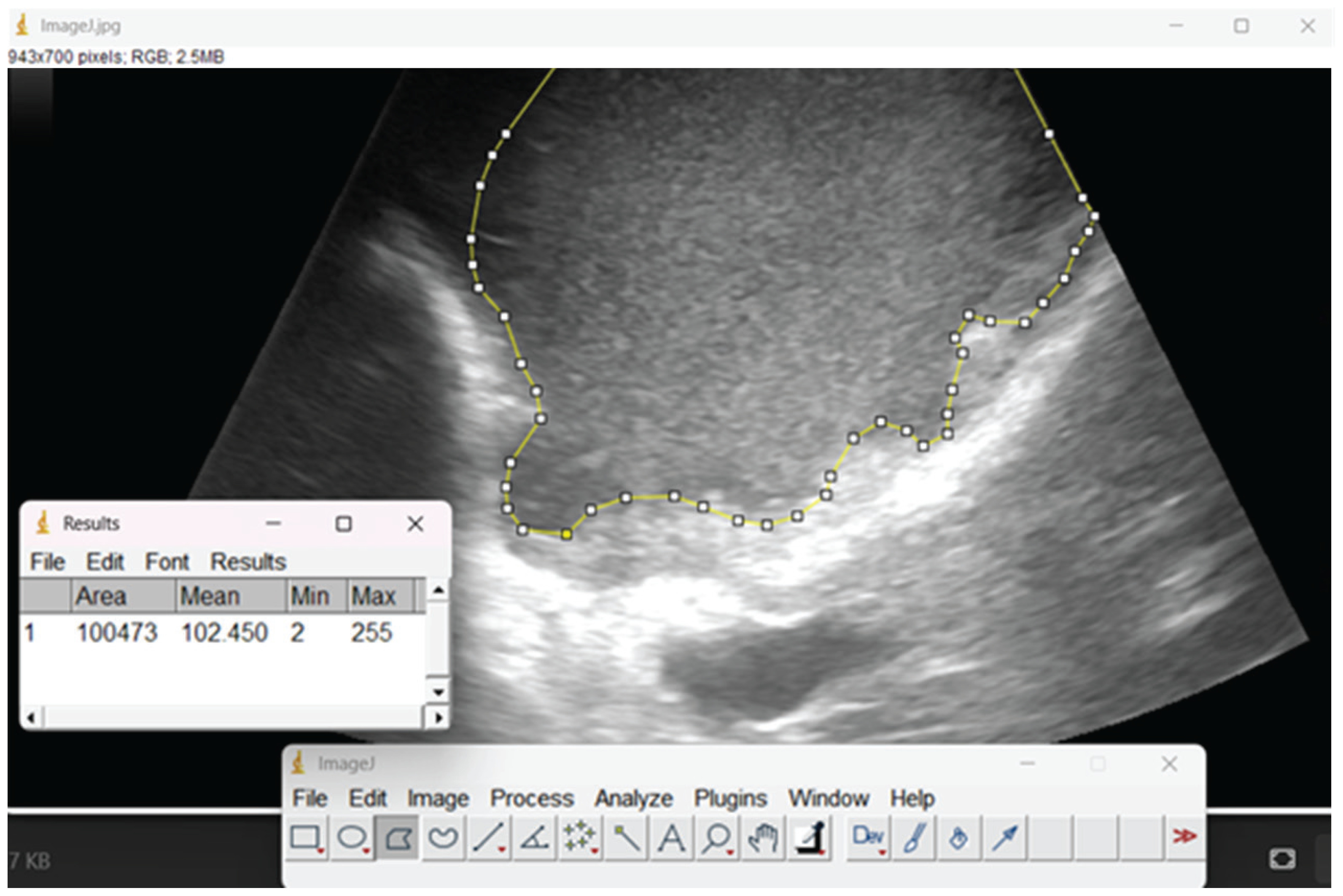
Using ImageJ software, a hypoechogenicity index – defined as the ratio of pleural fluid echogenicity to rib cortex – has been investigated, showing correlations with lactate dehydrogenase, cell count, and pH in parapneumonic effusions19.
In a separate retrospective cohort of 83 patients, pixel density quantification demonstrated significantly higher median values in exudates compared with transudates, achieving high specificity (>90%) at higher thresholds. A hybrid ultrasound scoring system combining pixel density with morphological features such as fibrin strands, debris, and loculations yielded positive predictive values exceeding 90%, although evidence remains exploratory and requires prospective validation20.
However, substantial technical limitations exist. Ultrasound machine settings dramatically affect measurements – minor gain adjustments can shift pixel density by >50 grayscale units. Measurements require manual region-of-interest (ROI) selection and labor-intensive ImageJ post-processing, impractical for bedside point-of-care assessment. Sensitivity remains disappointingly low (22–85%). Small, predominantly retrospective studies limit definitive conclusions. While conceptually interesting as a research tool, quantitative ultrasound cannot currently replace biochemical pleural fluid analysis and requires extensive multicenter validation with standardized protocols21.
Reproducibility represents a further critical constraint: the absence of a validated, standardized protocol for ROI delineation introduces substantial inter-observer variability. Without consensus on probe selection, gain settings, depth parameters, and ROI placement methodology, measurements from different operators or institutions are unlikely to be directly comparable, further limiting the clinical translatability of this approach.
Ultrasound elastography: investigating tissue characterization
Ultrasound elastography assesses tissue mechanical properties by quantifying the degree of deformation induced by an applied or intrinsic mechanical stimulus. Two principal techniques have been applied to pleural tissue: strain elastography, which estimates relative tissue compression under manual probe pressure, and shear wave elastography, which measures the propagation velocity of induced shear waves to derive quantitative stiffness values. Malignant pleural tissue typically exhibits greater stiffness than benign tissue, providing a biophysical rationale for differential diagnostic application.
Elastography allows non-invasive quantification of pleural stiffness, which is generally greater in malignant compared with benign effusions. Prospective data suggest this approach can achieve high diagnostic performance, with sensitivity and specificity reported around 84% and 91%, respectively, exceeding that of standard TUS22. Although it can help localize suspicious areas and inform biopsy targeting, elastography cannot provide a definitive diagnosis on its own.
In a multicenter cohort of 98 patients, most without significant pleural thickening or nodularity, elastography-guided biopsy yielded diagnoses in over 90% of cases and detected malignancy with nearly 90% sensitivity23. A subsequent randomized study in 218 patients confirmed improved detection in cases lacking overt pleural abnormalities, with sensitivity rising from 50% using conventional ultrasound to 80% with elastography guidance24.
Despite these promising results, routine adoption is constrained by operator dependence, inter-device variability, and the lack of standardized protocols25. Emerging evidence positions elastography-guided pleural biopsy as a less invasive alternative to thoracoscopy for patients with malignant effusions, negative cytology, and normal-appearing pleura on imaging. While thoracoscopy will likely remain the reference for tissue diagnosis, refinements in ultrasound-based techniques, particularly elastography, have the potential to enable safer, simpler, and more patient-focused strategies for managing malignant pleural effusions26.
Contrast-enhanced ultrasound: investigating biopsy optimization
Contrast-enhanced ultrasound (CEUS) uses gas-filled microbubble contrast agents to enhance real-time depiction of tissue perfusion and vascularity, allowing identification of viable, perfused tissue and helping to avoid necrotic areas when selecting a biopsy target. These agents remain intravascular and are cleared primarily via the lungs by exhalation of the gas component, making CEUS a repeatable technique that does not involve ionizing radiation and is not associated with nephrotoxicity.
In pleural-based lesions, a three-year prospective study of 460 patients demonstrated that CEUS detected internal necrosis in 72% of cases versus 32% with conventional ultrasound (p<0.05) and peripheral vessels in 55% versus 21% (p<0.05), achieving a diagnostic accuracy of 98.9% and markedly increasing biopsy yield in malignant (16% → 94%) and infectious lesions (77% → 100%)27.
More broadly, a systematic review and meta-analysis of 16 studies including 3459 thoracic and pulmonary lesions found that CEUS-guided biopsy consistently improved diagnostic performance compared with conventional ultrasound, with higher success rates (99.2% vs 97.3%), superior accuracy (96% vs 86%), and lower complication rates28. Collectively, these data indicate that CEUS-guided biopsy can substantially enhance diagnostic confidence for pleural and thoracic lesions.
However, limitations constrain routine implementation. CEUS requires specialized equipment, intravenous contrast with associated costs, and operator expertise. Its cost-effectiveness remains unproven. Importantly, most studies enrolled patients with radiologically apparent lesions; the benefit of CEUS in individuals without visible pleural abnormalities remains untested, representing a critical unmet need. Current evidence is largely derived from single-center studies, predominantly in Asian populations, and multicenter validation is lacking. Consequently, while CEUS-guided biopsy is a promising, minimally invasive approach for selected pleural lesions, its use should currently be considered adjunctive and context-dependent rather than routine.
Artificial intelligence: early promises and validation challenges
AI applications in respiratory medicine are increasingly explored for their potential to support clinical decision-making, optimize patient management, and improve diagnostic and prognostic accuracy29-32. In TUS, AI has generated considerable interest for its potential to reduce operator dependence and enhance diagnostic performance33.
A systematic review of five studies encompassing 7565 patients using convolutional neural networks for pleural effusion detection reported sensitivities of 71–100%, specificities of 67–100%, and AUC of 0.77–0.9934. For pneumothorax detection, systematic reviews found sensitivities of 79–94% and specificities of 80–87%35.
However, fundamental limitations constrain clinical implementation. All studies showed high risk of methodological bias, predominantly retrospective designs, and inadequate separation of training and testing data – leading to inflated performance estimates. Most critically, when tested on new patient populations from different centers, diagnostic accuracy dropped substantially, with sensitivity declining from >95% to 71–82%34. Models performed poorly on challenging cases and showed strong dependence on image quality. No studies have evaluated whether AI actually improves patient outcomes, alters clinical decisions, or provides cost-effective benefit. Current evidence remains insufficient for routine clinical use.
Notably, while these performance metrics appear promising in controlled settings, direct comparative data between AI models and experienced clinicians performing conventional TUS remain scarce. In external validation settings, AI sensitivity for pleural effusion detection declined substantially (71–82%)34, and trained operators may perform comparably or superiorly under real-world conditions – a critical benchmark that future prospective, head-to-head studies must explicitly address. Beyond methodological limitations, the development of robust AI tools for thoracic ultrasound requires access to large, well-annotated datasets encompassing diverse patient populations, imaging platforms, and acquisition conditions – a resource that remains limited. Regulatory approval, transparent decisionmaking frameworks, and seamless integration into clinical workflows represent additional prerequisites for clinical adoption that current studies have not yet adequately addressed36.
Research priorities and methodological requirements
Advancing these techniques toward validated clinical tools requires addressing several critical priorities. Prospective multicenter studies with consecutive enrolment are essential to generate realistic performance estimates across diverse populations, ultrasound platforms, and healthcare settings. Current evidence is largely derived from retrospective single-center studies with selected cohorts; future studies must deliberately include challenging cases rather than exclude them.
Urgent standardization is required. Scanning protocols, measurement methods, and diagnostic thresholds vary widely, precluding meaningful comparison. International consensus initiatives should define standardized acquisition parameters, quality metrics, and reporting frameworks for each technique, alongside structured training program to ensure operator competence.
Studies must assess clinically meaningful outcomes beyond diagnostic accuracy. Key questions include whether Doppler screening reduces bleeding complications, whether AI improves decision-making or efficiency, and whether advanced techniques influence treatment selection or patient-centered outcomes such as quality of life. Cost-effectiveness analyses are essential, and randomized controlled trials comparing these approaches with standard care remain the methodological gold standard.
For AI-specific applications, methodological rigor is paramount. Studies must enforce strict patient-level data separation, perform external validation on independent datasets, report transparently in accordance with TRIPOD-AI guidelines, include direct comparison with expert clinicians, and incorporate explainability frameworks demonstrating focus on clinically relevant features, verified through expert review36.
The potential complementarity and synergistic integration of the reviewed techniques merit explicit consideration in future research frameworks. Rather than being evaluated in isolation, M-mode functional assessment, quantitative pixel-based imaging, elastography, and CEUS may represent synergistic components of a multimodal ultrasound evaluation. AI-based analytical tools may ultimately provide a unifying framework capable of simultaneously processing data from multiple modalities – including elastography-derived stiffness maps and pixel-based echogenicity data – to support more comprehensive and reproducible diagnostic workflows. Future studies should explore how these complementary approaches can be rationally combined and sequenced in clinically meaningful diagnostic protocols.
Expert image review and peer validation through structured surveys encompassing multiple experienced operators should be considered as complementary strategies to improve reproducibility and build consensus on image interpretation standards. This approach is particularly relevant in ultrasound-based and AI-assisted diagnostics, where formal inter-observer agreement data remain limited and where the definition of reference standards is inherently dependent on operator expertise.
Finally, AI-based tools may serve not only as diagnostic adjuncts but also as educational instruments – supporting skill acquisition, standardizing training curricula, and potentially accelerating competence development among trainees and less experienced operators. Incorporating this perspective into the design of future training programs and competence assessment frameworks may broaden the impact of AI applications in thoracic ultrasound beyond diagnostic support alone.
Limitations
As a narrative review, this work is subject to several inherent limitations. The literature search and study selection were conducted without a pre-specified systematic protocol; the absence of a formal screening process introduces the possibility of selection bias, including potential under-representation of negative or null findings. Studies were prioritized based on clinical relevance, methodological quality, and recency, which may not fully reflect the breadth of available evidence. The evidence base for all techniques discussed remains limited by predominantly small, single-center, and retrospective studies; accordingly, conclusions must be interpreted with appropriate caution. These limitations are acknowledged throughout the text and reinforce the overarching recommendation that all reviewed techniques be regarded as investigational until validated by prospective, multicenter evidence incorporating rigorous clinical assessment and patient-centered outcomes.
CONCLUSION
Imaging in respiratory medicine is advancing at an unprecedented pace. Advanced TUS techniques and AI applications offer new opportunities to improve diagnostics, enhance safety, and reduce operator dependence. However, evidence remains limited, and these approaches are still investigational.
Clinicians should interpret current findings cautiously, and prospective multicenter studies incorporating rigorous clinical assessment, patient-centered outcomes, and cost-effectiveness analyses are needed to define true utility.
Until robust evidence is established, conventional TUS by trained operators, guided by sound clinical judgement, remains the cornerstone of pleural disease assessment. Any future evidence must be interpreted in the context of clinical expertise and patient-centered care, ensuring that emerging technologies complement – not replace – individualized assessment and decision-making.